|
|
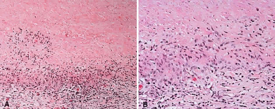
| Fig. 32. Palisading necrobiotic granuloma. A. The upper half of the field is hypocellular and consists of necrobiotic collagen. A palisade of histiocytes borders this, and a sparse lymphocytic infiltrate is present beyond at the bottom of the figure (hematoxylin and eosin, × 10). B. High power showing the layer of palisading histiocytes (hematoxylin and eosin, × 20). |