|
|
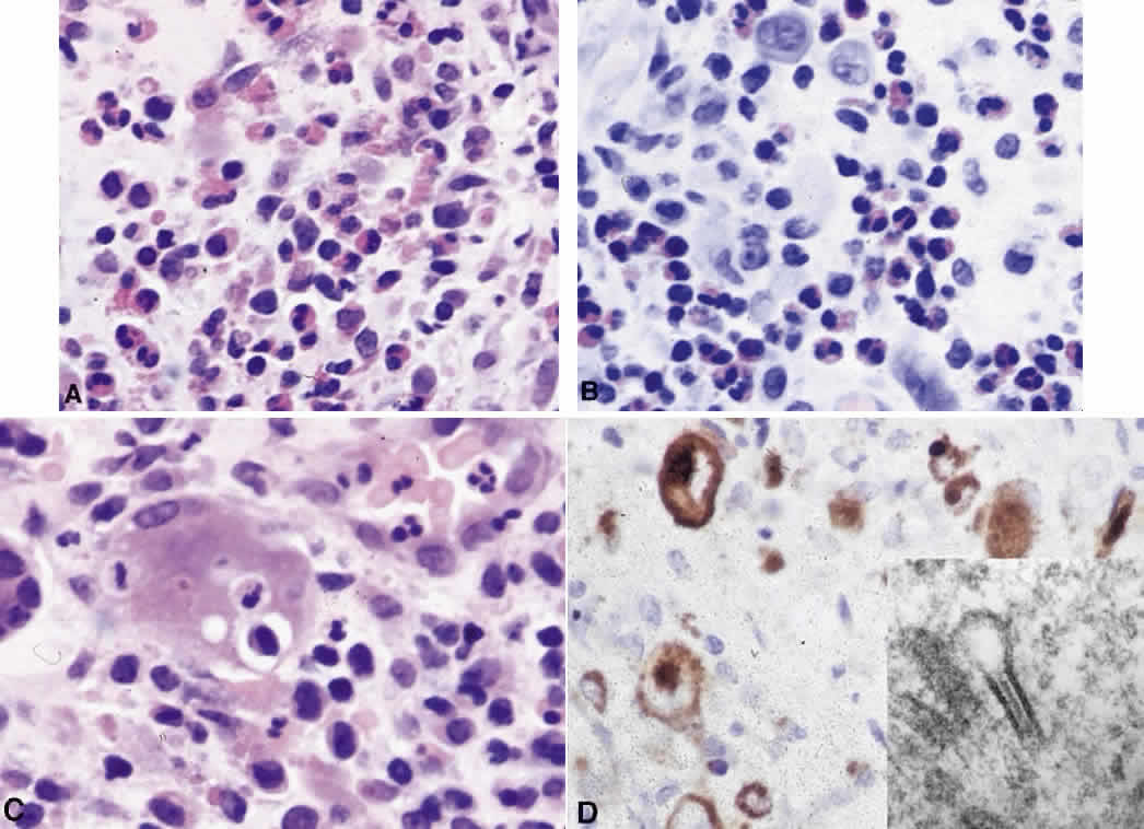
| Fig. 26. Langerhans' cell histiocytosis. A. High-power micrograph shoes the presence of numerous eosinophil polymorphs and histiocytes (hematoxylin and eosin, × 40). B. A section stained with Sirius red highlighting the eosinophil polymorphs within the lesion (Sirius red, × 40). Some of the histiocytes are mononuclear and contain “coffee bean”-like nuclei. C. Other histiocytes are giant cells containing phagocytosed debris (hematoxylin and eosin, × 40). D. Stain with peanut lectin agglutinin immunohistochemical examination. Langerhans' cells stain with a characteristic paranuclear dot and surface membrane pattern of staining (× 40). Insert: An electron micrograph of a “tennis racket”—shaped Birbeck granule (× 105). |