|
|
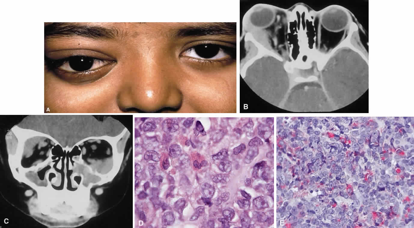
| Fig. 24. A. A 10-year old boy with advanced bilateral orbital chloroma evident clinically on the right. Axial (B) and coronal (C) CT scan of the orbit showing bilateral orbital and sinus involvement. D. Histologic examination shows the presence of occasional mature eosinophil granulocytes mixed with less mature myeloblast-like cells (hematoxylin and eosin, × 60). E. Chloroacetate esterase (Leder) staining confirms these cells to be of granulocytic type (Leder, × 40). (Courtesy of Dr. I. A. Cree) |