|
|
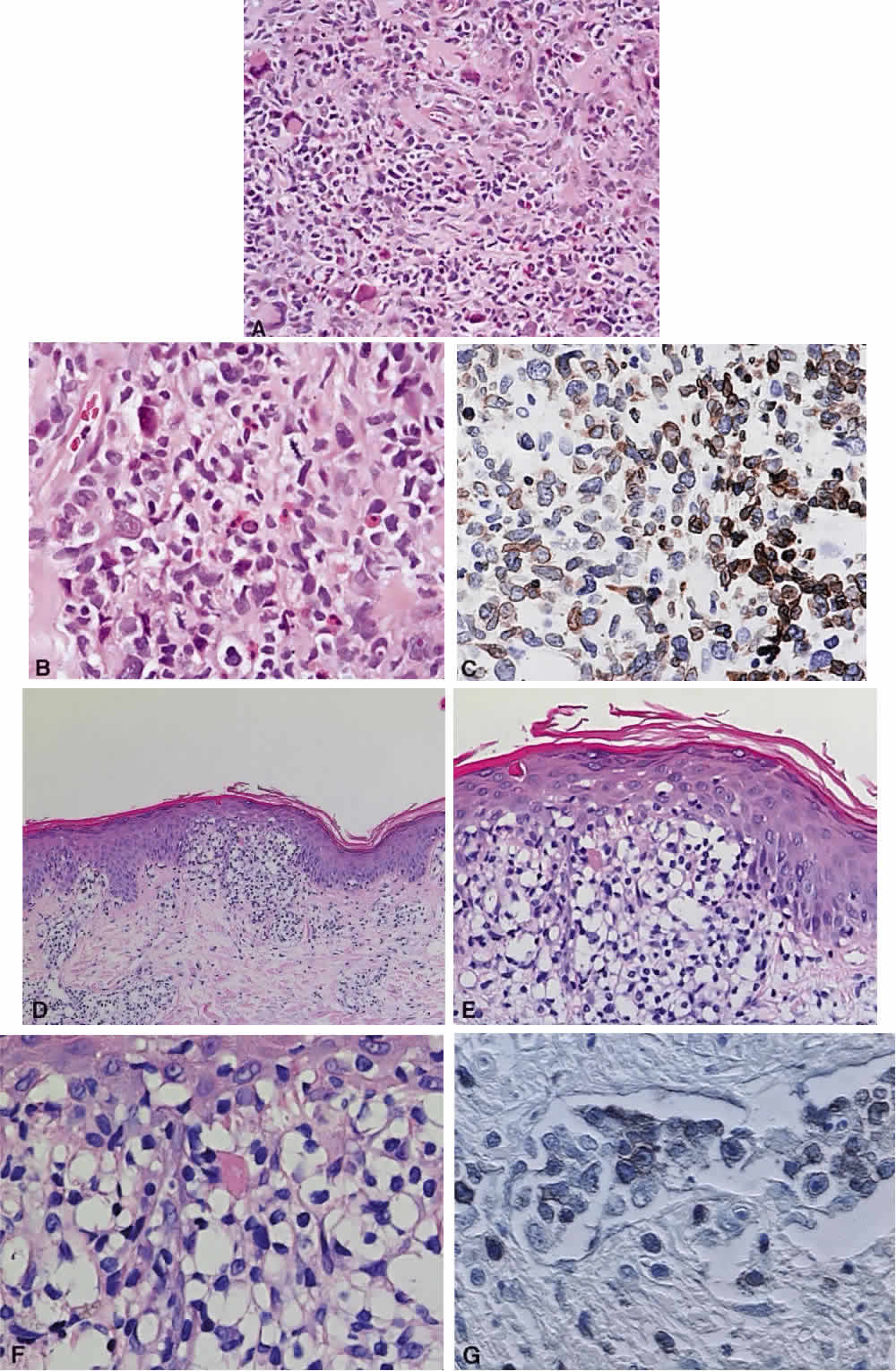
| Fig. 19. T-cell lymphomas. A. Peripheral T-cell lymphoma (unspecified): A mixture of small and medium-sized lymphoid cells with occasional large pleomorphic cells are a few eosinophils interspersed (hematoxylin and eosin, × 20). B. There is cellular pleomorphism with mitotic figures and a prominent blood vessel at top left (hematoxylin and eosin, × 40). C. CD3 immunophenotyping demonstrates the abnormal lymphoid cells to be of T-cell lineage (immunoperoxidase, × 40). D. Mycosis fungoides with a dermal lymphoid infiltrate showing epidermotropism at the dermal-epidermal junction and perivascular infiltration elsewhere (hematoxylin and eosin, × 10). E. Epidermotropism with the disruption of the normal dermal-epidermal border and infiltration of cells into the epidermis (hematoxylin and eosin, × 20). F. Higher power of the same lesion with a mixture of small and large lymphoid cells, some with irregular nuclei (hematoxylin and eosin, × 60). G. Immunophenotyping of the abnormal lymphoid cells using CD3 antibody, shows them to be of T-cell lineage (× 60). (Case courtesy of Dr. A. Mowat.) |