|
|
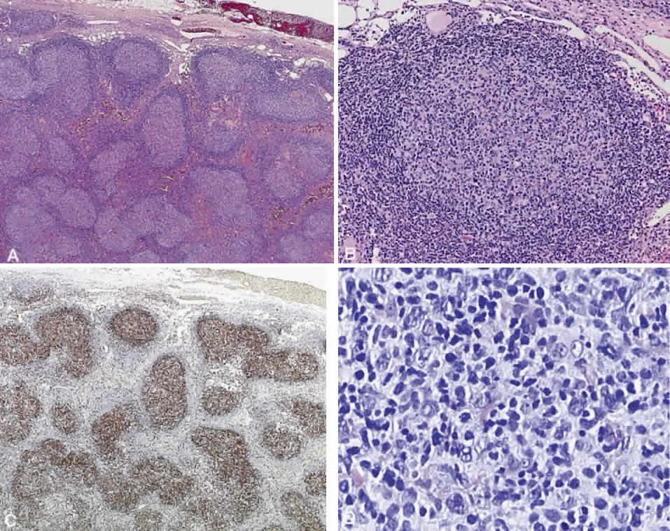
| Fig. 10. Follicle center cell lymphoma. A. Multiple abnormal follicles of variable size and shape with a surrounding mantle zone (hematoxylin and eosin, × 2). B. An abnormal follicle showing the absence of tingible body macrophages within the follicle cell center (hematoxylin and eosin, × 2). C. Antibody stain for bcl-2 highlights the centers of the abnormal follicles, a pattern typical of follicle center cell lymphoma (immunoperoxidase, × 2). D. Center of an abnormal follicle showing a mixture of pleomorphic cells with “cleaved” and “noncleaved” nuclei. Tingible body macrophages are absent (hematoxylin and eosin, × 40). |