|
|
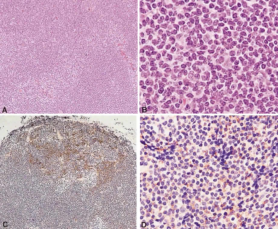
| Fig. 9. Extranodal marginal zone lymphoma. A. A diffuse infiltrate with ill-defined nodularity (hematoxylin and eosin, × 10). B. Cytologic features of the neoplastic lymphoid cells include small centrocyte-like cells, some with plasmacytoid nuclei (hematoxylin and eosin, × 60). C. CD21 stain shows the presence of preexisting lymphoid follicle, which has been partially destroyed by infiltration with marginal zone cells (immunoperoxidase, × 10). D. CD20 immunophenotyping demonstrates B-cell lineage (immunoperoxidase, × 40). |