|
|
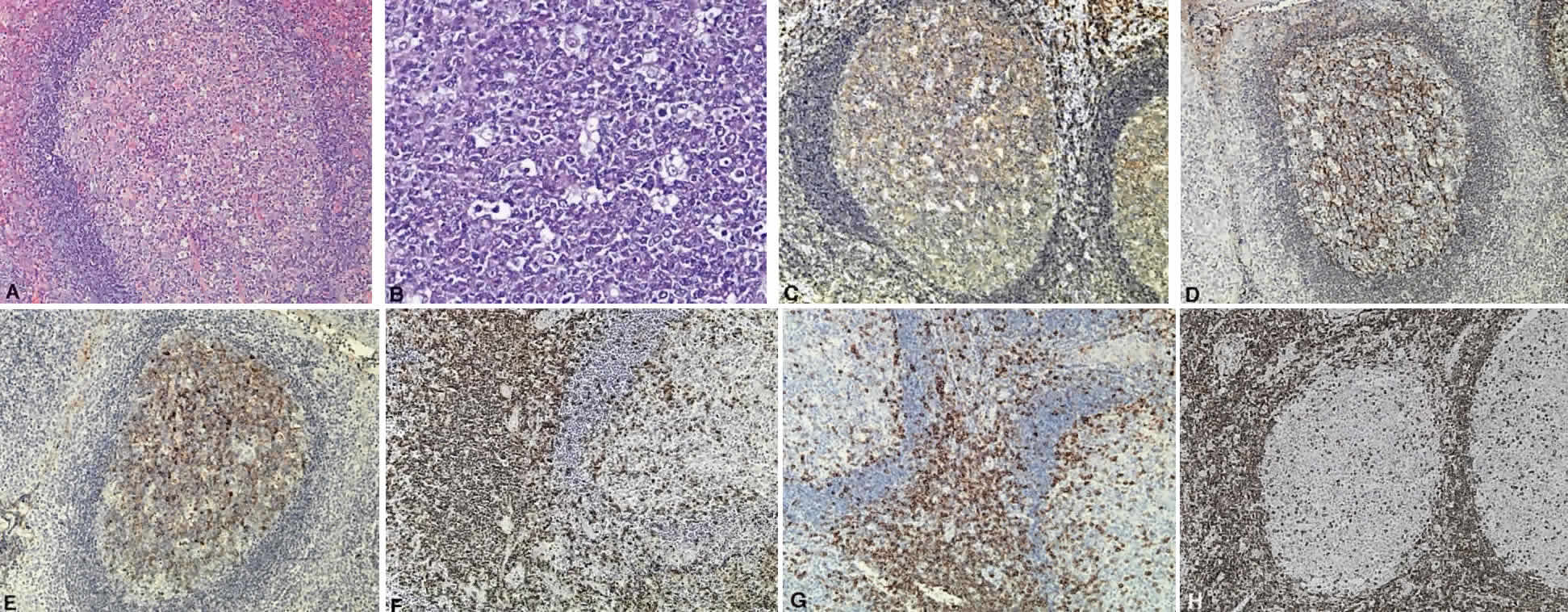
| Fig. 2. The normal lymphoid follicle. A normal follicle is round or ovoid with a pale center containing tingible body macrophages. A. A well-defined zone of small dark lymphocytes surrounds the follicle separating it from the interfollicular zone (hematoxylin and eosin, × 10). B. A follicle center showing the follicle center cells with “cleaved” or “noncleaved” nuclei interspersed with tingible body macrophages (hematoxylin and eosin, × 20). C. CD20 demonstrates the follicle center cells, the mantle zone cells, and many of the interfollicular cells to be B cells (immunoperoxidase, × 10). D. CD21 staining illustrates the meshwork of dendritic follicular cells that provide a scaffold for the follicle center cells (immunoperoxidase, × 10). E. Follicle center cells express CD10 (immunoperoxidase, × 10). F. T cells are found in the interfollicular zone with few penetrating the follicle, as demonstrated by CD3 staining (immunoperoxidase, × 10). G. T cells express CD5 and also are predominantly found in the interfollicular zone, whereas some penetrate the follicle (immunoperoxidase, × 10). The oncoprotein bcl-2 is expressed normally by mantle zone and some interfollicular zone cells. H. Few cells of the follicle center express bcl-2, in contrast to follicle center cell lymphoma (immunoperoxidase, × 10). |