|
|
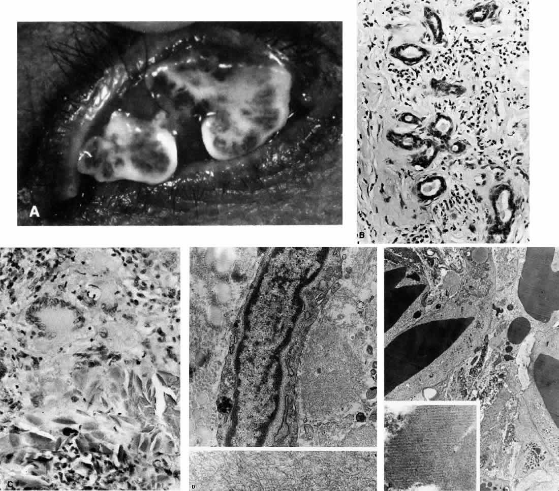
| Fig. 9. Amyloidosis. A. Recurrent conjunctival vegetations with mild chronic inflammation and tremendous amyloid content. B. Amorphous intercellular material has replaced most of the lacrimal gland, with few glandular units surviving. There is an infiltrate of chronic inflammatory cells, many of which are plasma cells (H&E, 74% of ×240). C. Amorphous eosinophilic material, giant cell, epithelioid cells, and fragments of needle-like particles, which have elicited foreign body reaction, are shown. The condition is termed tissue proteinosis (H&E, ×240). D. Electron micrograph from a case of lacrimal gland amyloidosis shows a surviving cell, its identity unascertainable, with cytoplasmic filaments and profiles of rough endoplasmic reticulum, surrounded on right by amyloid fibrils and on left by collagen. There is an osmiophilic inclusion at the bottom left of the cell. In the lower panel, amyloid material is seen as slender (10 nm wide) nonbranching fibrils (Top, ×8000; bottom, ×25,000). E. Osmiophilic material with crystalline substructure (shown in inset) is being engulfed by degenerating giant cells and macrophages, which have abundant cytoplasmic filaments (F). It is possible that intracytoplasmic filaments, many of which may have been extended into the intercellular space after dissolution of cell membranes, are responsible for borderline amyloid stain. Lattice-like material is proteinaceous, with resemblance to crystallized immunoglobulin (×5,000; inset, ×60,000). |