|
|
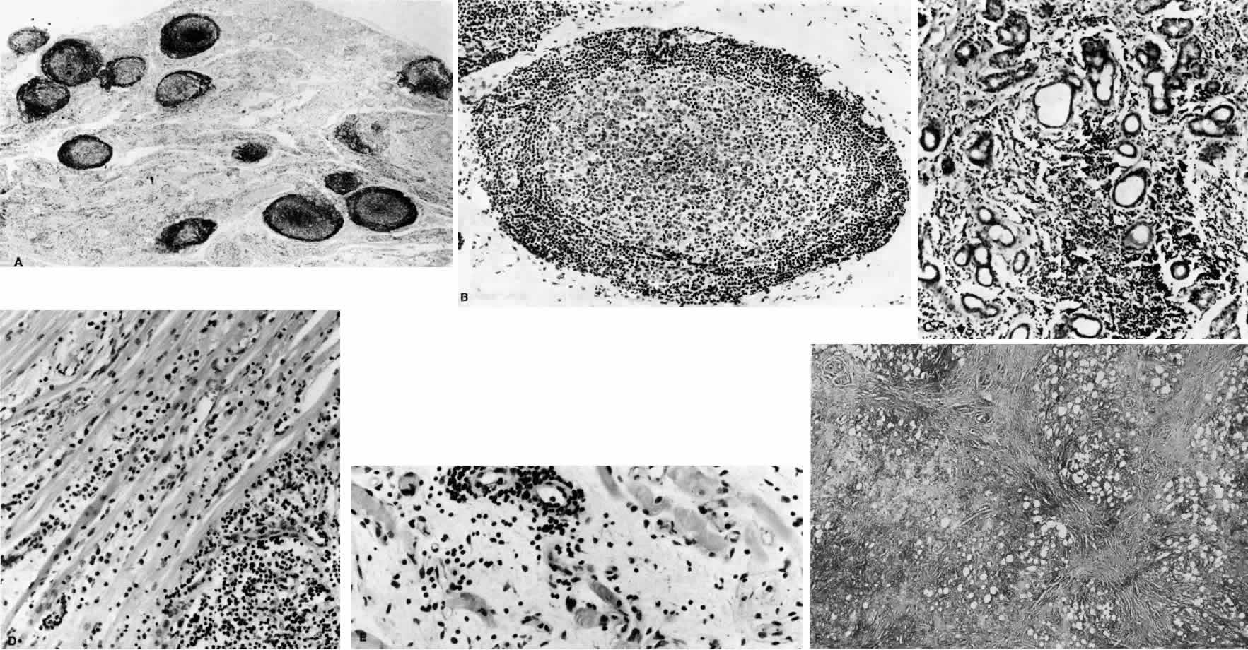
| Fig. 6. Histopathologic sections demonstrating the various pathologic findings associated with idiopathic orbital inflammation. Most commonly these include lymphoid follicles, granulomas, collagen deposition, and a diffuse mixed inflammatory cell infiltrate. The normal tissue architecture is frequently disrupted by these changes. A. Many lymphoid follicles are scattered throughout orbital tissue (H&E, × 63). B. Cells of follicular center are lighter and larger than mantle of mature lymphocytes that surround the germinal zone (H&E, ×160). C. Lacrimal gland elements have undergone atrophy in advanced example of idiopathic dacryoadenitis. Fibrosis and lymphocytes have replaced a considerable amount of gland parenchyma (H&E, ×94). D. Myositis in which lymphocytes are loosely aggregated below center and infiltrate between extraocular muscle fibers (H&E, ×160). E. Cuffing of small vessels by mature lymphocytes. Note loose edematous interstitium between disrupted muscle fibers (H&E, ×240). F. Progressive fibrosis of retrobulbar fat (H&E, ×25). |