|
|
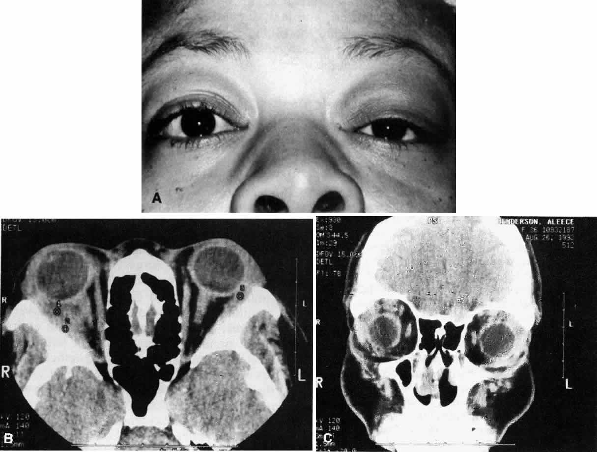
| Fig. 1. A. A 36-year-old woman presented with right-sided pain and proptosis occurring over a several-day period. Externally the eye is quiet and does not appear inflamed. The patient had no complaints of diplopia or other visual disturbances. Tissue obtained at biopsy demonstrated idiopathic orbital inflammation. B. Axial CT image from the same patient demonstrates bilateral orbital masses located laterally in the orbits. Note that masses are ill defined and do not appear to have a capsule. The masses are molded to the bony orbital walls without evidence of any bony irregularities. C. Coronal CT image from the patient again shows the presence of bilateral orbital masses apparently involving the lateral rectus muscles. |