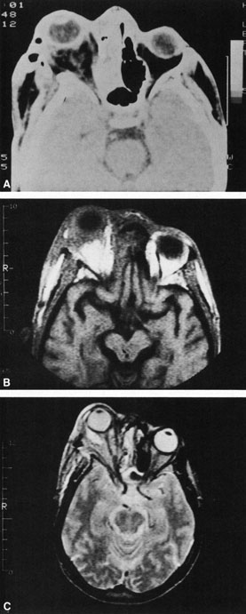
Fig. 18.
Orbital abscess.
A.
Computed tomography of an orbital abscess presenting as an enhancing intraconal mass on right side.
B.
T1-weighted image.
C.
T2-weighted image. Note area of high signal corresponding to abscess.