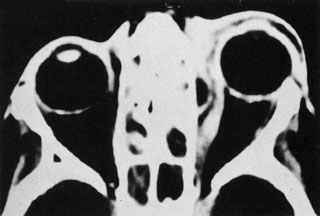
Fig. 16.
Computed tomography showing subperiosteal abscess formation. Note elevation of orbital periosteum and convexity as pus elevates periorbit from the medial orbital wall.