|
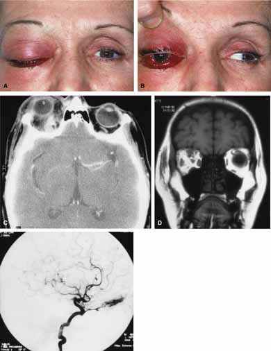
Fig. 11
A. A 48-year-old female presented with sudden proptosis (developing over several minutes), pain, ptosis, decreased ocular motility, decreased visual acuity, and conjunctival chemosis. B. Conjunctival chemosis and restricted motility is seen. C. On axial computed tomography (CT) scanning a contrast enhancing mass is seen superiorly. D. Coronal magnetic resonance imaging (MRI) scanning revealed the superior anomaly. E. Angiography demonstrated an arteriovenous malformation of the right orbit. The vascular malformation was successfully removed surgically.
|