|
Fig. 9
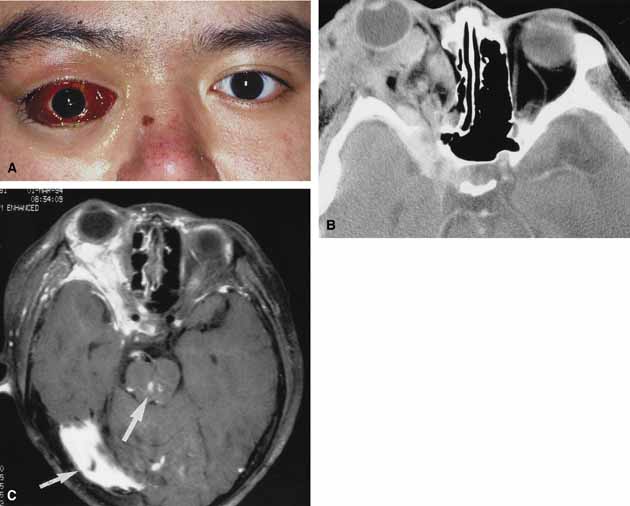
A. A 20-year-old male with extensive right facial and orbital lymphangioma presented acutely with proptosis, subconjunctival hemorrhage, double vision, and orbital pain. B. Axial computed tomography (CT) scan reveals extensive involvement of the orbit. The abnormal tissue also extends through the superior orbital fissure to the cavernous sinus area. C. Magnetic resonance imaging (MRI) scan reveals not only the extensive orbital involvement but also vascular anomalies in the midbrain and cerebellum (arrows).
|