|
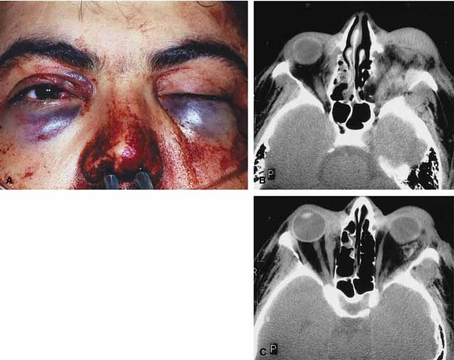
Fig. 7
A. A 20-year-old male presented to the emergency department after a knife attack with bilateral periocular hemorrhages, ptosis, and proptosis of the left eye; normal vision on the right; and a vision of 20\/200 on the left. Both globes were intact. B. Axial computed tomography (CT) scan with signs of orbital hemorrhage bilaterally (Lt > Rt). C. Axial computed tomography (CT) scan with signs of orbital hemorrhage bilaterally (Lt > Rt).
|