|
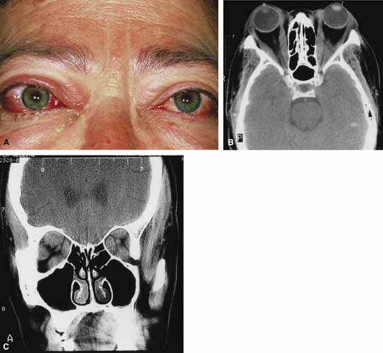
Fig. 6
A. A 56-year-old female presented with a 2-day history of proptosis, lid swelling, conjunctival chemosis/injection, and double vision. Thyrotropin (TSH) level was extremely low and a computed tomography (CT) scan revealed grossly enlarged extraocular muscles. A diagnosis of acute thyroid eye disease was made. B. Axial computed tomographt (CT) scan illustrating diffuse enlarged extraocular muscles characteristic of thyroid eye disease. C. Coronal computed tomography (CT) scan showing enlargement of extraocular muscles.
|