|
|
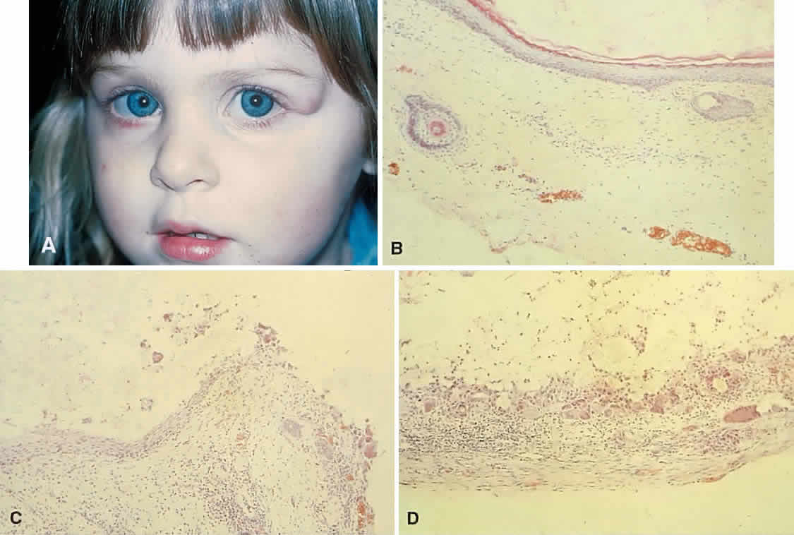
| Fig. 19. A. This 2-year-old girl had a mass overlying the superotemporal orbital rim since birth. The area had enlarged and had become red and tender in the preceding few weeks. B. The recent clinical changes are explained by rupture of the cyst wall and a granulomatous inflammatory response to the expelled contents. This section shows typical stratified squamous epithelium, hair shafts in the wall, and keratin in the lumen. C. Transitional zone between the dermoid cyst wall and an encapsulated granulomatous response. D. The wall of the cyst beyond the point of rupture. Note the multinucleated giant cells and fibrous capsule. (B-D, hematoxylin-eosin; × 96.) |