|
|
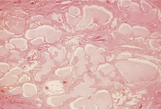
| Fig. 11. The labyrinthine structure of a lymphangioma appears as multiple microcysts in histologic sections. The lumens contain pale staining lymphlike fluid. The channels extend into surrounding normal tissue without circumscription or encapsulation (hematoxylin-eosin; × 40). |