|
|
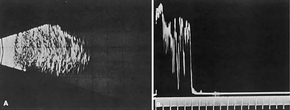
| Fig. 9. A. B-scan echography shows marked internal acoustic heterogeneity and lack of circumscription. B. Quantitative A-scanning shows a corresponding pattern. High-amplitude spikes are reflected from the vessel-lumen/cell-cluster interfaces within the tumor. |