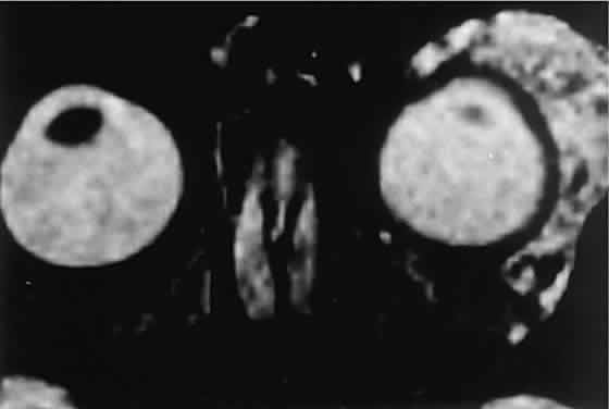
Fig. 8.
Gadolinium-enhanced, fat-suppressed, T
2
-weighted MRI shows the extent of a large capillary hemangioma that infiltrates the lacrimal gland. Dark streaks represent flow voids within feeding and draining vessels.