|
|
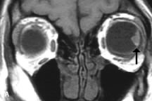
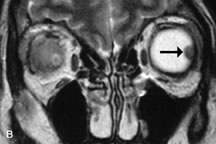
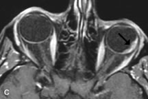
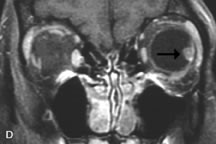
| Fig. 24. A. T1- and (B) T2-weighted MR scans demonstrate a small nodular intraocular mass (arrows) that is very hyperintense on the T1-weighted scan and hypointense on the T2-weighted image. This signal intensity pattern is due to the presence of free radicals within melanin granules. C and D. Postcontrast fat-suppressed T1-weighted scans demonstrate homogeneous intense enhancement of the lesion and no evidence of seleral penetration or optic nerve invasion. |