|
|
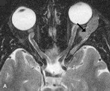
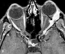
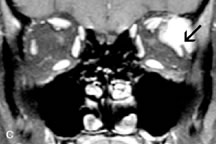
| Fig. 23. A. T2-weighted and (B and C) postcontrast fat-suppressed T1-weighted MR scans demonstrate an infiltrative lacrimal region mass than invades the lateral rectus muscle (arrows). This highly cellular lesion is seen to have a very hypointense appearance on the T2-weighted scan. |