|
|
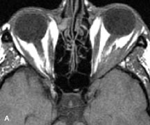
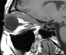
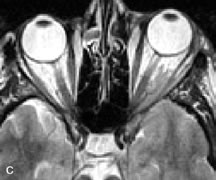
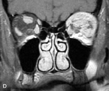
| Fig. 22. A and B. T1- and (C) T2-weighted MR scans demonstrate a poorly defined multicompartmental mass enveloping the lateral rectus, superior rectus, and levator palpebrac superioris muscles. The lesion is isointense to brain on T1- and T2-weighted scans, as is typical for highly cellular neoplasms. D. Postcontrast fat-suppressed T1-weighted scan demonstrates intense enhancement of the infiltrating intraconal and extraconal tumor. |