|
|
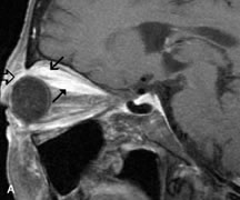
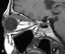
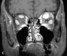
| Fig. 21. A. T1-weighted MR scan demonstrates diffuse enlargement of both the superior rectus and levator palpebrae superioris muscles (single arrows). The involvement of the tendinous insertions and preseptal soft tissues (open arrows) as well as lack of involvement of other muscles helps differentiate this entity from thyroid-associated orbitopathy. B and C. Postcontrast fat-suppressed T1-weighted MR scans demonstrate extensive enhancement of the involved muscles as well as the preseptal (open arrows) and perinuscular tissues (double arrows). |