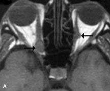
Fig. 20.
A.
T1-weighted MR scan demonstrates nodular enlargement of both medial rectus muscles (
arrows
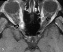
). B. T1-weighted fat-suppressed contrast-enhanced scan confirms the presence of small metnstatic deposits within the muscles (
open arrows
).