|
|
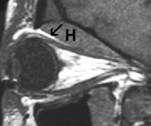
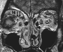
| Fig. 19. A. T1- and (B) T2-weighted MR scans demonstrate a large acute subperiosteal hematoma (H) that lies between the cortical bone of the orbital roof and the inferiorly displaced periorthira (double arrow). The extracopal fat (arrow) and levator muscle are displaced inferiorly. |