|
|
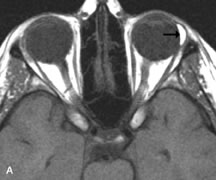
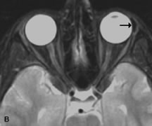
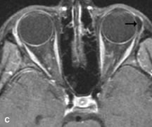
| Fig. 18. A. T1-weighted, (B) T2-weighted fat-suppressed, and (C) T1-weighted fat-suppressed MR scans demonstrate a small dermoid cyst arising near the palpebral portion of the lacrinal gland (arrows). The lesion is similar in signal intensity to fat on the T1-weighted scan (A) consistent with a high adipose tissue content. The lesion shows fat-suppression and low signal intensity on the two fat-suppressed sequences (B and C) confirming its high lipid content. |