|
|
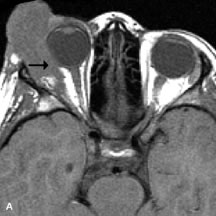
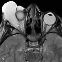
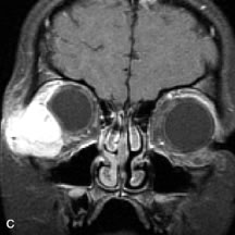
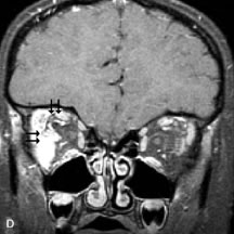
| Fig. 17. A. T1- and (B) T2-weighted MR scans demonstrate a preseptal and extraconal mass displacing the globe medially. The mass is slightly hyperintense on the T1-weighted scan and very hyperintense on the T2-weighted scan owing to the high extracellular water content of the neoplasm. The lesion abuts the globe and appears to infiltrate the lateral rectus muscle (arrow). C and D. Postcontrast fat-suppressed T1-weighted scans demonstrate intense enhancement of the highly infiltrative lesion that is invading the lateral rectus, superior rectus, and levator palpebrac superioris muscle (double arrows). |