|
|
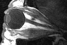
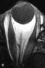
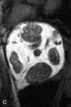
| Fig. 16. A-C, T1-weighted MR scans obtained with a high-resolution surface coil demonstrate fusiform enlargement of the extraocular muscles. The medial, lateral, and inferior rectus muscles are especially involved. Note the relative sparing of the tendinous insertions, a finding characteristic of this disease process, as well as fatty infiltration of the lateral and inferior rectus muscles. There is marked proptosis, best visualized on the sagittal image (A), and mild crowding of the optic nerve at the orbital apex. |