|
|
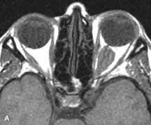
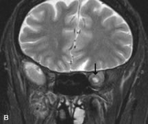
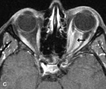
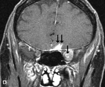
| Fig. 15. A. T1- and (B) T2-weighted MR scans demonstrate a mass causing fusiform enlargement of the optic nerve sheath complex. The peripheral portion of the mass, representing the meningioma, is isointense to brain on the T1-weighted scan and notably hypointense on the T2-weighted scan. The central portion of the mass, representing an edematous optic nerve, is very hyperintense on the T2-weighted scan (arrow), C and D. Postcontra fat-suppressed T1-weighted scans demonstrate intense peripheral enhancement of the meningioma surrounding the central nonenhancing optic nerve (arrow). Also note the enhancing Intracranial “dural tail” (double arrow). |