|
|
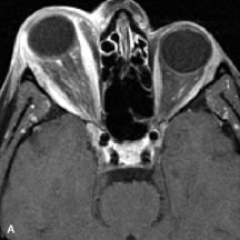
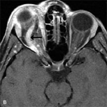
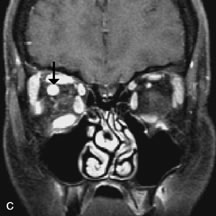
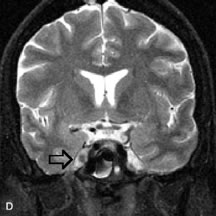
| Fig. 14. Patient with Ehlers-Danlos syndrome who presented with abrupt onset of severe proptosis. A-C. Postcontrast fat-suppressed T1-weighted MR scans demonstrate marked proptosis and engorgement of the extraocular muscles and superior ophthalmic vein (arrows). D. T2-weighted scan through the cavernous sinus demonstrates enlargement and arterialized flow void within the right cavernous sinus (open arrow). |