|
|
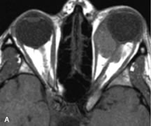
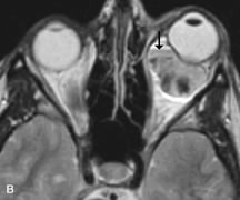
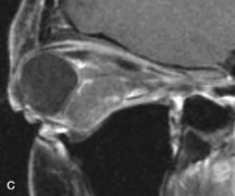
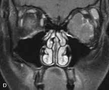
| Fig. 13. A. T1- and (B) T2-weighted MR scans demonstrate a moderately well-circumscribed intraconal mass enveloping the optic nerve. The lesion is hyperintense to vitreous on the T1-weighted scan. A fluid-fluid level (arrow) and patchy hypointense areas are seen within the mass on T2-weighted scan due to the presence of subacute blood products. C and D. Postcontrast fat-suppressed T1-weighted scans demonstrate no significant enhancement within the lesion, although there is minimal peripheral enhancement in surrounding orbital tissues. |