|
|
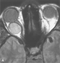
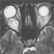
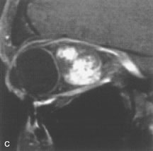
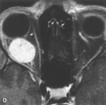
| Fig. 12. A. T1- and (B) T2-weighted MR scans demonstrate a well-circumscribed intraconal mass causing severe optic nerve displacement. These lesions are usually intermediate in signal intensity on T1-weighted scans and very hyperintense on T2-weighted Images. C and D. Postcontrast fat-suppressed T1-weighted scans demonstrate characteristic patchy intense enhancement that becomes more complete from the initial postcontrast scan (C) to a more delayed scan (D). |