|
|
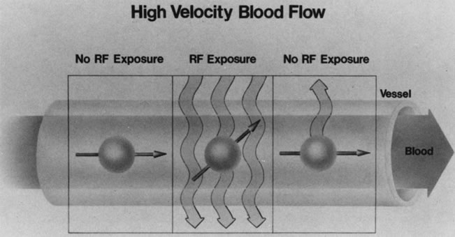
| Fig. 10. The lumina of blood vessels containing rapidly flowing blood appear dark because the excited protons in the blood leave the imaging slice before the emitted signal can be detected. This phenomenon is illustrated by dividing a blood vessel into three frames. The nucleus in the central frame energized by the radiofrequency (RF) signal passes out of the imaging plane into the right frame before it relaxes and emits a signal. In the left frame, the hydrogen nucleus is not exposed to the RF waves and therefore cannot give a signal as it enters the imaging plane in the central frame. Therefore, no signal is detected in the blood vessel, which consequently appears dark, (Dortzbach RK, Kronish JW, Gentry LR: Magnetic resonance imaging of the orbit, Part I, Physical principles. Ophthal Plast Reconstr Surg 5:156, 1989) |