|
|
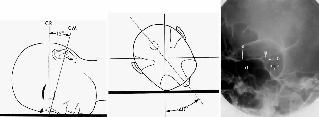
| Fig. 6. A and B. Schematic showing positioning for an oblique apical projection (Rhese position). (CR, central ray; CM, canthomeatal line) C. Radiograph of an oblique apical projection. (a, right optic canal; b, optic strut; c, superior orbital fissure; d, ethmoid sinus; e, planum sphenoidale; f, greater wing of sphenoid) (A and B; Rao VM, Gonzalez CF: Plain film radiography and polytomography of the orbit. In Gonzalez CF, Becker MH, Flanagan JC [eds]: Diagnostic Imaging in Ophthalmology, pp 1–7. New York, Springer Verlag, 1986) |