|
|
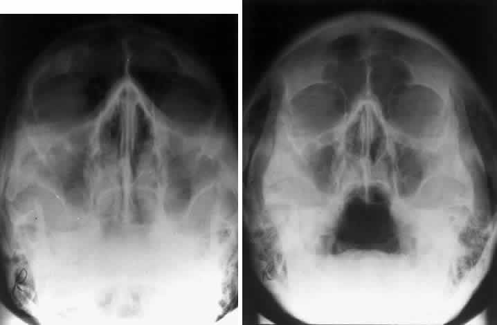
| Fig. 1. In any projection the image detail is maximized using the minimal distance from the subject and the x-ray cassette. A. Anterior to posterior projection demonstrating decreased clarity and definition of anterior structures. B. Posterior to anterior projection showing the improved clarity of the anterior structures, such as the orbital rim and frontal sinus. |