|
|
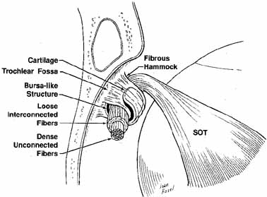
| Fig. 16 Schematic drawing of the right trochlea. Tendon is supported by a layer of cartilage suspended by fibrous supports from the periorbita. Central fibers of the tendon are strong with dense unconnected fibers. Peripheral tendon shows loose interconnected fibers. (SOT, superior oblique tendon) (Adapted from Helveston EM, et al: The trochlea: A study of the anatomy and physiology. Ophthalmology 1982:89:124) |