|
|
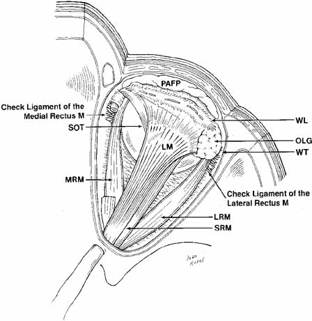
| Fig. 11 Superior view of the orbit. Whitnall's ligament fuses medially with the trochlea of the superior oblique muscle and fuses laterally with the lacrimal gland. The medial horn of the levator aponeurosis lies directly on top of the superior oblique reflected tendon. The lateral horn of the levator aponeurosis splits the palpebral and orbital lobe of the lacrimal gland. The lateral rectus check ligament attaches to Whitnall's tubercle and is slightly denser than the medial rectus ligament. (WL, Whitnall's ligament; OLG, orbital lobe of lacrimal gland; SOT, superior oblique tendon; PAFP, preaponeurotic fat-pad; LM, levator palpebrae superioris muscle; WT, Whitnall's tubercle; MRM, medial rectus muscle; LRM, lateral rectus muscle; SRM, superior rectus muscle) |