|
|
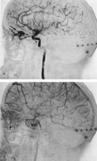
| Fig. 33. Orbital arteriovenous fistula. A 58-yearold man noted redness of the left eye, blurred vision, and diplopia 3 years following head trauma, which had resulted in brief unconsciousness and amnesia for 2 hours. The left eye was initially red but recovered fully, and the patient was asymptomatic in the interval. Examination showed proptosis, slight limitation of motion of globe, and bruit over orbit. A: Left carotid arteriogram demonstrates vascular mass in the roof of the left orbit (arrow) with marked arteriovenous shunting from the ophthalmic artery. B: Venous phase shows choroidal crescent of the globe (arrow), slightly flattened and displaced anteriorly. Surgical excision at the University of California, San Francisco, was successful, with disappearance of all signs and symptoms. (Courtesy of Dr. Gene Coin.) |