|
|
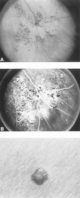
| Fig. 29. Cavernous hemangioma of the retina. Fundus appearance (A) and fluorescein angiogram (B) of segmental angiomatous malformation of retina. Note saccular dilation of capillary shunts with pooling of dye. C: Cutaneous angioma in same patient. (Courtesy of Dr. J.D.M. Gass.) |