|
|
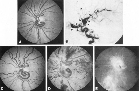
| Fig. 28. Wyburn-Mason syndrome. A 4-year-old child with left amblyopia, acuity 20/200 (6/60). A: Dilated venous anomaly, left disc. B: Subtracted left internal carotid angiogram shows both an arteriovenous malformation along course of ophthalmic artery (arrows) and a suprasellar vascular anomaly (arrowheads). C: Progressive enlargement of venous complex. D: Within 20 months, vision reduced to finger-counting, the fundus appearance was that of hemorrhagic ischemic retinopathy, and neovascular angle-closure glaucoma developed with applantation tension of 54 mm Hg. Panretinal xenon laser photocoagulation was performed. E: Repeat fluorescein angiography shows clearing of retinopathy and diminished perfusion of anomalous vessels. Applanation tension was reduced to 10 mm Hg. (Effron L, Zakov ZN, Tomsak RL. Neovascular glaucoma as a complication of the Wyburn-Mason syndrome. J Clin Neuro Ophthalmol 5:95, 1985) |