|
|
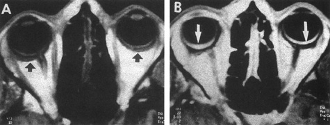
| Fig. 26. Sturge-Weber disease. Axial magnetic resonance imaging sections. A: T1-weighted axial image shows thickening of posterior aspect of both globes (arrows). B: T1-weighted image with contrast shows bilateral diffuse choroidal hemangiomas (arrows). (From ref. 138) |