|
|
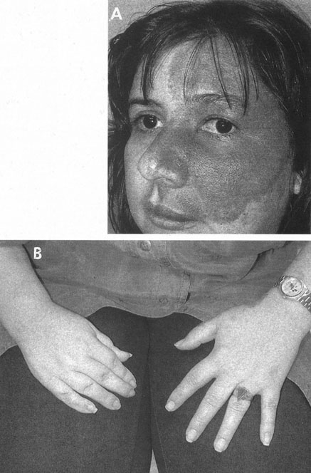
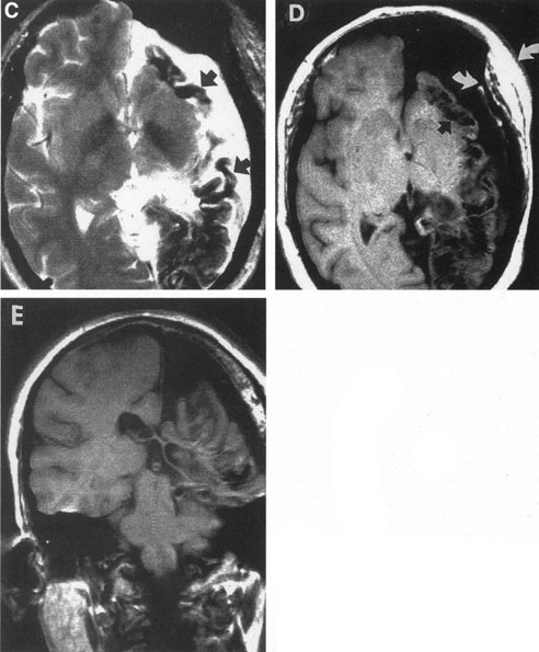
| Fig. 24. Sturge-Weber disease. A 34-year-old woman with life-long seizures and with right homonymous hemianopia. A: Verrucous facial nevus flammeus; note that cutaneous angioma in this case precisely outlines right ophthalmic and maxillary divisions of the trigeminal nerve. B: Relative somatic hypoplasia on the right (small right hand) owing to a left cerebral vascular lesion. C: T2-weighted magnetic resonance imaging (MRI) shows hemicerebral atrophy, thickening of meninges caused by venous angioma, and calcification (black areas, arrows) that occurs in outer layers of cortex and in meningeal arteries. D: T1-weighted magnetic resonance imaging (MRI). Note calcified gyri (black areas) and compensatory thickening of overlying cranium (arrows). E: MRI, coronal section. |