|
|
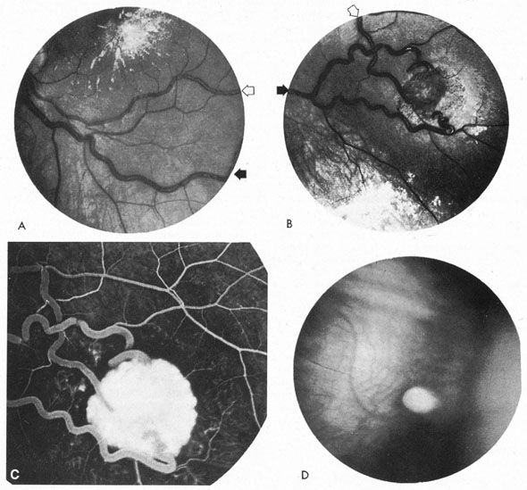
| Fig. 23. Angiomatosis retinae (von Hippel's tumor). A: A 23-year-old man presented with diminished vision from exudative retinopathy involving the left macula. B: Typical retinal angioma was noted in inferotemporal midperiphery. Open arrows indicate feeding retinal arteriole; closed arrows indicate draining vein. C: Fluorescein angiogram demonstrates arteriovenous shunt through angioma. D: Small berry-like angioma in far retinal periphery. (A through C, Courtesy of Dr. J.D.M. Gass.) |