|
|
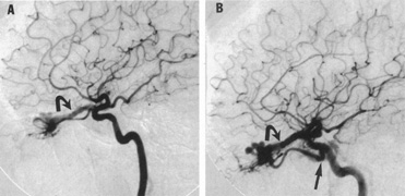
| Fig. 19. A 30-year-old woman presented with headaches. Lateral views of left (A) and right (B) internal carotid arteriograms demonstrate developmental venous malformation of the cribriform plate supplied by both ophthalmic arteries. Note subarachnoid veins of the anterior cranial fossa (curved arrows). In addition, the right ophthalmic artery (B) has a cavernous origin (arrow), a vestige of the dorsal ophthalmic artery that usually atrophies in fetal development. |