|
|
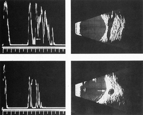
| Fig. 16. Orbital ultrasonography. Transocular A-scans (left) and B-scans (right) in a carotid–cavernous fistula with dilated arterialized superior ophthalmic vein. Top left: Blurred spikes (arrow) within the dilated vessel indicate fast blood flow. Bottom left: Distinct spikes (arrows) from vessel walls, at low system sensitivity. Top right. Dilated superior ophthalmic vein (arrow), at low sensitivity setting. Bottom right: Cross-section of enlarged superior ophthalmic vein. (From Byrne SF, Glaser JS: Orbital tissue differentiation with standardized echography. Ophthalmology 90:1071, 1983.) |