|
|
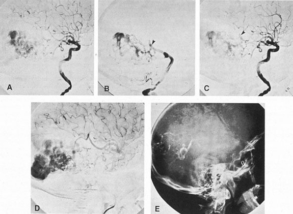
| Fig. 11. Embolization of middle cerebral vessels that supply occipital lobe arteriovenous malformation (AVM). The patient had a subarachnoid and intraparenchymal hemorrhage that produced a left homonymous field defect. A: Right carotid arteriogram demonstrates contribution via posterior communicating artery to a right occipital lobe AVM. B: Vertebral injection. The arrow points to the enlarged right posterior cerebral artery that is a major feeder of the AVM. C: Right carotid arteriogram during glue embolization procedure. The arrow points to a catheter as it traverses the segment seen in (B). The catheter was advanced via the internal carotid artery but is positioned far posteriorly. D: Upper branches to the AVM now are occluded, with residual low-flow vascularization via the middle cerebral artery. E: Skull film showing radiopaque glue within the AVM and blood vessels previously supplying it. The patient had a persistent visual field defect but greatly reduced headache and no persistence of subjective bruit. (Courtesy of Dr. Joseph Horton.) |