|
|
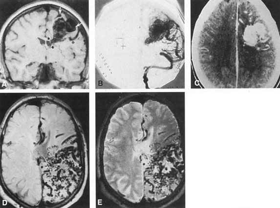
| Fig. 10. Neuroimaging of arteriovenous malformation. A: Coronal magnetic resonance imaging (MRI) (TR, 600 ms; TE, 20 ms) shows wedge-shaped vascular mass extending from the surface of the parietal cortex (white arrows) to the lateral ventricle (black arrows; asterisk, arterial feeder). B: Posteroanterior left arteriogram of the same lesion. C: Axial computed tomography scan with contrast enhancement. D: Axial MRI of a large arteriovenous malformation, first echo (TR, 2000 ms; TE, 20 ms). E: Second echo (TR, 2000 ms; TE, 90 ms). (From Smith HJ, Strother CM, Kikuchi Y, et al: MR imaging in the management of supratentorial intracranial AVMS. AJNR Am J Neuroradiol 9:225, 1988.) |