|
|
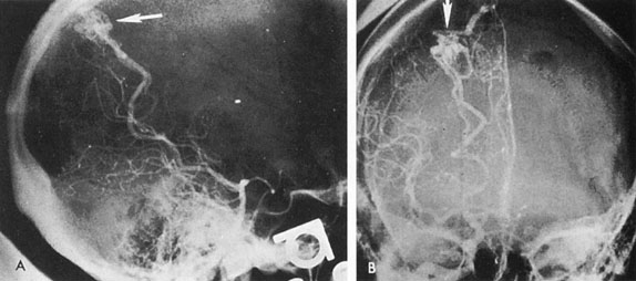
| Fig. 9. Carotid arteriogram of an occipital lobe arteriovenous malformation (AVM). Lateral (A) and frontal (B) projections demonstrating a small occipital AVM (arrow). The patient was a 23-year-old woman who presented with severe apoplectic unilateral headache, total left homonymous hemianopia, and mild nuchal rigidity. Despite xanthochromic cerebrospinal fluid, she was initially diagnosed elsewhere as having migraine. An AVM was successfully resected, and a small occipital lobe hematoma was removed. |