|
|
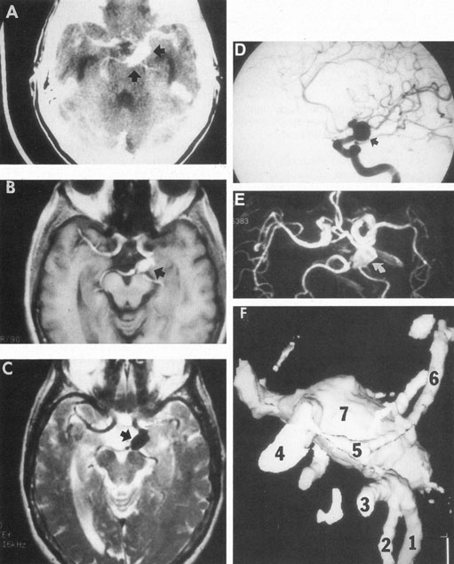
| Fig. 5. Aneurysm at the junction of the internal carotid and posterior communicating arteries. A: Contrast-enhanced axial computed tomography (CT) shows a large aneurysm (arrows). Magnetic resonance imaging (MRI) studies effectively demonstrate the aneurysm. B: T1-weighted MRI with gadolinium. C: T2-weighted (arrow indicates black flow void). D: Selective internal carotid angiogram. E: Magnetic resonance angiogram (MRA; curved arrow) shows the same aneurysm. F: Helical image-intensified CT technique provides quasi-three–dimensional mold of arteries: 1. right posterior cerebral; 2. posterior superior cerebellar; 3. basilar; 4. internal carotid; 5. posterior communicating; 6. left posterior cerebral; 7. supraclinoid aneurysm, same as depicted in A through E. (Courtesy of Dr. Raphael Aponte.) |